
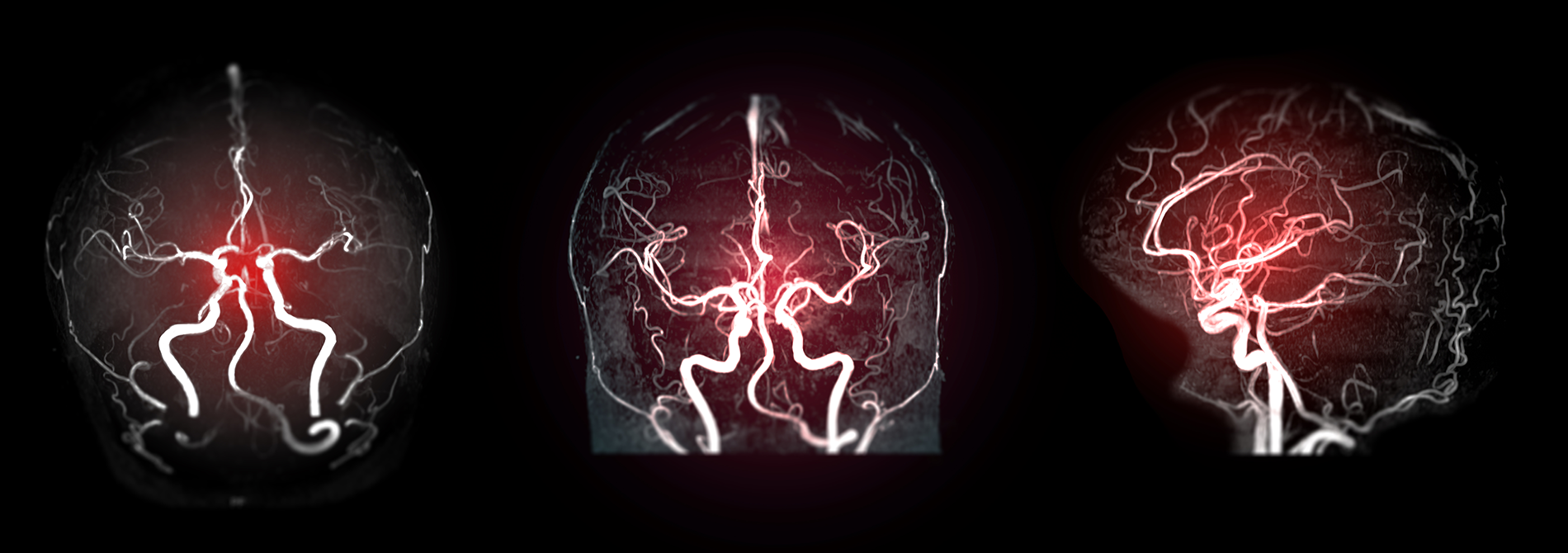
MRI angiography is a non-invasive imaging technique used to visualize blood vessels throughout the body. It uses magnetic resonance imaging (MRI) technology to create detailed images of blood vessels without the need for contrast dye injections or invasive procedures. MRI angiography can provide information about the size, shape, and condition of blood vessels, helping to diagnose conditions such as aneurysms, stenosis (narrowing), or vascular malformations. It is often used to evaluate the blood supply to organs and tissues, detect abnormalities in blood vessels, and plan treatments such as surgery or angioplasty.
Many patients with arterial disease now have it treated in the radiology department rather than undergoing surgery in an operating room. MRA is a very useful way of finding problems with blood vessels and determining how to best to treat those problems.
The carotid arteries in the neck that conduct blood to the brain are a common site of atherosclerosis, which may severely narrow or block off an artery, reducing blood flow to the brain and even causing a stroke. If an ultrasound study shows that such disease is present, many surgeons will perform the necessary operation after confirmation with MRA, dispensing with the need for catheter angiography.
MRA has found wide use in checking patients for diseased intracranial (in the head) arteries, so that only those with positive findings will need to undergo a more invasive catheter study.
MRA is also used to detect disease in the aorta and in blood vessels supplying the kidneys, lungs and legs.
Patients with a family history of arterial aneurysm, a ballooning out of a segment of the vessel wall, can be screened with MRA to see if they have a similar disorder that has not produced symptoms. If an aneurysm is found, it may be eliminated surgically, possibly avoiding serious or fatal bleeding.
• Bulges in your aorta, called aneurysms
• Tears in your aorta, called dissections
• Problems with your heart that you may be born with, called congenital heart disorders
• Narrowing of the arteries in and around your kidneys, called renal artery stenosis
• Inflammation in your blood vessels, called vasculitis
• Hardening of the arteries (called atherosclerosis) involving the legs or arms
• Blockages in the major arteries that supply blood to your brain, called carotid artery disease
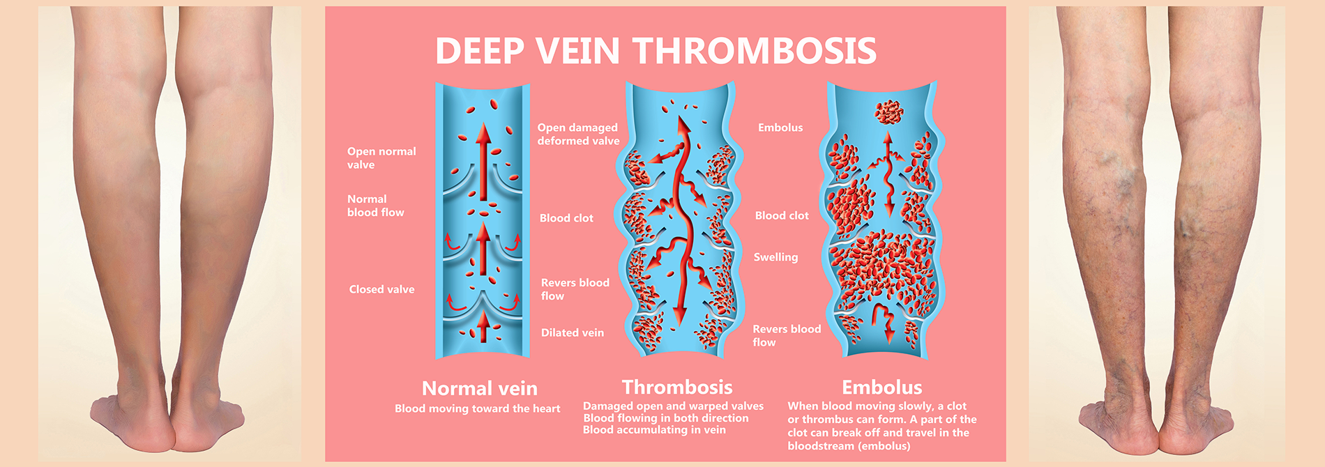
MRI venography is a type of magnetic resonance imaging (MRI) technique used to visualize veins in the body. It involves the use of MRI technology to create detailed images of the veins, typically without the need for contrast dye injections. MRI venography can provide information about the structure and function of veins, helping to diagnose conditions such as deep vein thrombosis (DVT), venous insufficiency, or varicose veins. It is particularly useful for evaluating blood flow within the venous system and identifying any abnormalities or blockages that may be present.
Two common techniques to obtain the images of flowing blood are:
• TOF – Time of flight: Incoming blood makes the vessels appear bright, and surrounding tissue is suppressed.
• SSFP – Steady state free precession is generally used for assessment of veins in the chest, abdomen, and pelvis.
Although the initial evaluation of the iliac and lower extremity veins is usually accomplished with Ultrasound. MRV is more efficient in detecting venous thrombosis in the pelvic and calf veins.
Please setup a login with Clearpath here:
Why are we using Clearpath?